
This lab explores the stages of mitosis by observing onion root tip cells under a microscope. It helps students understand cell division and its significance in growth and development.
1.1 Purpose of the Experiment
The purpose of the experiment is to observe and identify the stages of mitosis in onion root tip cells. By examining actively dividing cells under a microscope, students can visualize the different phases of the cell cycle, including interphase, prophase, metaphase, anaphase, and telophase. This hands-on activity helps students understand the process of cell division and its importance in plant growth and development. It also allows for the calculation of the time spent in each phase, providing insights into the dynamics of mitosis.
1.2 Importance of Studying Mitosis in Onion Root Tips
Studying mitosis in onion root tips is crucial for understanding plant growth and development. These cells are ideal due to their rapid division and accessibility. Observing mitosis helps students grasp fundamental biological processes and the cell cycle’s role in tissue repair and regeneration. This experiment bridges theoretical knowledge with practical observation, enhancing comprehension of cellular dynamics and their implications in biology.

Materials and Equipment Needed
Essential materials include onion bulbs, microscope, slides, cover slips, forceps, razor blade, staining dye, and water. These tools facilitate optimal observation of mitotic stages.
2.1 List of Required Materials
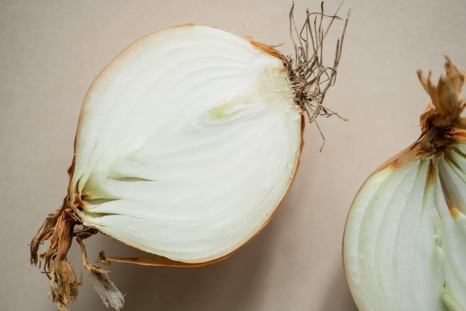
The materials needed include onion bulbs, a compound microscope, glass slides, cover slips, a sharp razor blade or scalpel, forceps, a staining dye like methylene blue or Toluidine Blue, distilled water, paper towels, and a clean workspace. These items are essential for preparing and observing the onion root tip cells effectively under the microscope to study the stages of mitosis. Proper preparation ensures clear visibility of cellular structures during the experiment.
2.2 Preparation of the Microscope
Before use, the microscope is cleaned with lens paper to remove dust and smudges. The stage is adjusted to hold the slide securely, and the focus knobs are set to align the lenses. The light source is adjusted for proper illumination. Start with the low-power objective lens to locate the cells, then switch to high power for detailed observation. Proper preparation ensures clear and accurate viewing of the onion root tip cells during the experiment.

Procedure for Observing Mitosis
Prepare the onion root tip section, stain it, and observe under a microscope to identify the stages of mitosis, ensuring clear cell visibility for accurate phase identification.
3.1 Preparing the Onion Root Tip Section
To prepare the onion root tip section, cut the root tip and fix it in water to stop cell division. Section the root thinly using a razor blade or microtome, ensuring transparency for staining. Mount the sections on slides and stain with acetoorcein or methylene blue to enhance chromosome visibility. This process preserves the cells and allows for clear observation of mitotic stages under a microscope, making it easier to identify and study each phase effectively.
3.2 Staining the Cells for Better Visibility
Staining is essential for enhancing the visibility of cell structures. Acetoorcein or methylene blue are commonly used to stain onion root tip cells. Gently heat the slide with the stain to help it penetrate the cells. After staining, rinse the slide with distilled water to remove excess dye. This process highlights the chromosomes and cell nuclei, making it easier to distinguish between the stages of mitosis under a microscope. Proper staining ensures clear observation and accurate identification of mitotic phases.
3.3 Observing the Cells Under the Microscope
After preparing and staining the root tip section, place it under the microscope. Start with low magnification (100x) to locate the meristem, where active cell division occurs. Once located, switch to high magnification (400x) for detailed observation. Focus on identifying cells in different mitotic phases, such as interphase, prophase, metaphase, anaphase, and telophase. Record observations systematically, noting the characteristics of each phase. Properly stained and prepared slides ensure clear visibility of chromosomes and cell structures, aiding accurate identification and analysis.

Phases of Mitosis
Mitosis consists of five distinct phases: interphase, prophase, metaphase, anaphase, and telophase. Each phase has unique characteristics essential for cell division, ensuring genetic continuity.
4.1 Interphase
Interphase is the longest phase of mitosis, where the cell grows, replicates its DNA, and prepares for division. During this phase, the chromatin remains uncoiled, and the nucleolus is visible. The cell increases in size, producing organelles and proteins essential for cell division. This phase is divided into three sub-stages: Gap 1 (G1), Synthesis (S), and Gap 2 (G2). DNA replication occurs in the S phase, ensuring each daughter cell receives identical genetic material.
4.2 Prophase
During prophase, chromatin condenses into visible chromosomes, and the nucleolus disappears. Spindle fibers form outside the nucleus, attaching to centrioles at the cell poles. The nuclear envelope disintegrates, allowing chromosomes to move toward the cell equator. This phase is critical as it prepares the cell for metaphase by organizing the chromosomes for alignment. The chromosomal structures become distinct, making them visible under a microscope, and the spindle fibers position themselves to facilitate chromosome separation in the subsequent stages of mitosis.
4.3 Metaphase
Metaphase is characterized by the alignment of chromosomes at the cell equator, attached to spindle fibers. Each chromosome is connected by microtubules to opposite poles. This phase ensures equal distribution of chromosomes to daughter cells. The chromosomes are tightly packed and visible under a microscope. Metaphase is critical for ensuring genetic material is divided accurately. Observing this phase helps in understanding how cells maintain genetic integrity during division. It is a brief but vital stage before anaphase begins.
4.4 Anaphase
Anaphase begins when sister chromatids separate, pulled to opposite poles by spindle fibers. This ensures each daughter cell receives an identical set of chromosomes. The nuclear envelope begins to disintegrate, and chromosomes appear to move rapidly. Anaphase is the shortest phase of mitosis, lasting only a few minutes. It is critical for ensuring genetic material is evenly distributed. Observing anaphase helps confirm the proper segregation of chromosomes, essential for cell division accuracy and genetic continuity. This phase sets the stage for telophase and cytokinesis.
4.5 Telophase
Telophase marks the final stage of mitosis, where the nuclear envelope reforms, and chromosomes uncoil into chromatin. The spindle fibers disappear, and the cytoplasm prepares for division. Each daughter cell now contains a complete set of chromosomes. This phase signals the transition to cytokinesis, where the cell splits into two. Telophase ensures the genetic material is organized for the next cell cycle, completing the division process. Observing telophase highlights the restoration of nuclear structure and the readiness for new cell formation.
Calculating the Time Spent in Each Phase
Estimate phase duration by counting cells in each stage and comparing proportions. Interphase occupies most time, while shorter phases like prophase and telophase take less time.
5.1 Method for Estimating Phase Duration
Count the number of cells in each phase under the microscope. Use a tally system to record observations. Calculate the proportion of cells in each phase by dividing the number of cells in a phase by the total number of cells observed. Multiply this proportion by the estimated duration of the cell cycle (typically 24 hours for plant cells). This provides an estimate of the time spent in each phase. Note that this method assumes cells are in a representative sample and may not account for variations in cell cycle duration. Some sources suggest using mitotic indices for more accurate calculations, while others recommend observing multiple root tips to ensure reliable data. This approach helps visualize and quantify the relative time cells spend in each stage of mitosis, reinforcing theoretical concepts with practical observations.
5.2 Interpreting the Results
After counting cells in each phase, compare the proportions to understand the duration spent in each stage. Most cells are in interphase, indicating it is the longest phase. Fewer cells in prophase, metaphase, anaphase, and telophase reflect their shorter durations. This aligns with cell cycle theories, where interphase dominates. Calculate the estimated time per phase by applying proportions to the total cell cycle duration (e.g., 24 hours for plant cells). These results highlight the dynamic nature of mitosis and its role in growth and tissue repair, providing practical insights into cellular biology.

Results and Data Analysis
This section presents the data collected from observing onion root tip cells, detailing the number of cells in each phase of mitosis and their percentages.
6.1 Recording Observations
Students observe onion root tip cells under a microscope, noting the stages of mitosis. They record the number of cells in each phase (interphase, prophase, metaphase, anaphase, telophase). Data is typically organized in a tally chart or table. Photographs or sketches of cells in different stages are often included for reference. Observations are meticulously documented to ensure accuracy and clarity for further analysis. This step is crucial for understanding the distribution of cells across mitotic phases and calculating phase durations.
6.2 Analyzing the Data
After recording observations, students analyze the data by calculating the percentage of cells in each phase of mitosis. This involves dividing the number of cells in a specific phase by the total number of cells observed and multiplying by 100. The results are compared to theoretical expectations, providing insights into the cell cycle. This analysis helps determine if cells spend equal time in each phase and deepens understanding of mitotic division patterns and their biological significance.
Drawing Conclusions
The experiment successfully demonstrated the stages of mitosis in onion root tip cells. Most cells were in interphase, confirming its prolonged duration; This supported theoretical cell cycle knowledge and enhanced understanding of mitotic division.
7.1 Summarizing the Findings
The experiment revealed that most onion root tip cells were in interphase, confirming its prolonged duration. A smaller percentage of cells were observed in prophase, metaphase, anaphase, and telophase. This distribution aligns with the understanding that interphase occupies the majority of the cell cycle. The findings also highlighted the challenge of capturing cells in the shorter, more dynamic phases of mitosis. The use of staining and microscopy effectively demonstrated the stages of cell division, supporting theoretical knowledge of mitotic activity in plant cells.
7.2 Relating the Results to Cell Cycle Theories
The lab results align with cell cycle theories, confirming that interphase is the longest phase, while prophase, metaphase, anaphase, and telophase are shorter. The distribution of cells across these stages supports the concept of continuous, regulated cell division. Observing the root tip cells under a microscope provided practical evidence of theoretical models, reinforcing the understanding of mitosis as a highly organized process essential for plant growth and development.
Pre-Lab and Post-Lab Questions
This section provides pre-lab questions to assess prior knowledge and post-lab questions to evaluate understanding of mitosis in onion root tips, ensuring comprehensive learning.
8.1 Pre-Lab Questions and Answers
What are the stages of mitosis in order?
⎯ Interphase, prophase, metaphase, anaphase, telophase.
Why are root tips used for observing mitosis?
⎯ Root tips have rapidly dividing cells, making them ideal for studying mitosis.
What is the purpose of using a microscope?
⏤ To observe and identify the stages of mitosis in onion root tip cells.
How do you prepare the root tip for observation?
⏤ Soak in water, fix, stain, and section.
Why is staining important?
⎯ It helps differentiate cellular structures under the microscope.
These questions ensure students are prepared for the experiment and understand its objectives.
8.2 Post-Lab Questions and Answers
What was the most challenging part of identifying mitosis stages?
⏤ Distinguishing between similar phases like prophase and metaphase;
Which phase did most cells appear to be in?
⎯ Interphase, as it lasts the longest.
How did staining improve observations?
⏤ It enhanced visibility of cell structures under the microscope.
What did you learn about the cell cycle?
⏤ Cells spend most time in interphase and less in division phases.
Why is this experiment important?
⎯ It demonstrates mitosis and its role in growth and tissue repair.
These questions reinforce understanding of mitotic processes and experimental outcomes.

Common Mistakes and Troubleshooting
- Improper fixation can distort cells, making mitosis stages hard to identify.
- Over-staining may obscure cell details under the microscope.
- Failure to focus correctly can lead to blurry observations.
9.1 Identifying and Avoiding Errors
- Improper fixation of root tips can distort cell structures, making mitosis stages hard to identify. Use fresh samples and follow fixation protocols carefully.
- Over-staining can obscure cell details. Use the recommended staining time to maintain visibility of cellular structures.
- Poor focus and incorrect microscope settings can lead to blurry observations. Ensure the microscope is properly calibrated before use.
- Contamination during slide preparation can ruin samples. Handle materials sterilely and avoid touching the slide surface.
- Rushing the procedure may result in missed mitotic phases. Examine multiple fields of view thoroughly.
9.2 Tips for Improving Observations
- Use high-magnification lenses (400x) for clearer cell visibility and detailed phase identification.
- Ensure proper staining to enhance contrast between cell structures and the background.
- Prepare thin, uniform root tip sections to avoid overcrowded cell layers.
- Examine multiple fields of view to increase the chances of observing all mitotic phases.
- Use reference images or diagrams to compare and confirm the stages of mitosis accurately.
Additional Resources and References
- Marietta College Introductory Biology Lab Manual provides detailed mitosis observation protocols.
- Onion Root Tip Mitosis Lab Guide offers practical tips and images for identifying cell stages.
- Onion Root Tip Mitosis Lab Report PDF includes answers and reference materials for students.
10.1 Recommended Reading Materials
For deeper understanding, refer to Marietta College Introductory Biology Lab Manual, which provides detailed protocols for observing mitosis. The Onion Root Tip Mitosis Lab Guide offers practical tips and images for identifying cell stages. Additionally, Cell Biology: A Short Course by Eliane Eltzroth gives a comprehensive overview of mitosis. These resources are essential for preparing and completing the lab report effectively, ensuring accurate observations and analysis of cellular processes.
10.2 Online Resources for Further Study
Visit biologyeducation.org for interactive guides and images of onion root tip cells in mitosis. Khan Academy’s cell biology section offers detailed explanations of mitotic phases. Additionally, labmanual.org provides downloadable guides for mitosis labs, including answer keys and protocols. For visual learning, explore virtual labs on platforms like virtuallab.org, which simulate the onion root tip experiment and offer step-by-step analysis tools.
The onion root tip mitosis lab effectively demonstrates cell division stages, providing insights into mitosis and its role in plant growth, enhancing understanding of cellular biology fundamentals.
11.1 Summary of Key Takeaways
The onion root tip mitosis lab highlights the importance of studying cell division in rapidly dividing cells. Observing mitosis under a microscope helps visualize its stages: interphase, prophase, metaphase, anaphase, and telophase. The root tip is ideal due to its high cell division activity. Staining techniques enhance cell visibility, allowing accurate phase identification. Calculating phase durations reveals interphase as the longest. This lab reinforces understanding of mitosis and its role in growth, providing practical experience in cellular biology.
11.2 Final Thoughts on the Experiment
The onion root tip mitosis lab provides valuable hands-on experience in understanding the cell cycle. Observing mitosis firsthand bridges textbook concepts with practical observation. Challenges like identifying phases accurately and avoiding cell damage highlight the need for meticulous technique. This experiment underscores the importance of mitosis in growth and development, reinforcing biological principles. It serves as a foundational skill for future studies in cellular biology and genetics.